
Panoramic Dental X-ray

What is a panoramic dental X-ray?

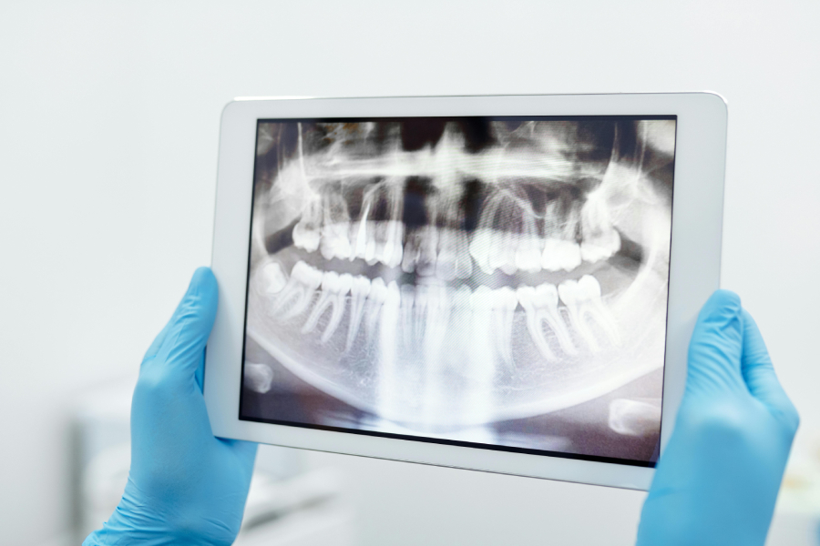
The panoramic dental X-ray or orthopantomography is an X-ray that provides a complete image of the oral cavity, covering both jaws and the teeth in a single image.

This examination is used to assess the overall condition of the teeth, jaws, and surrounding tissues, providing valuable information about the health of the oral cavity. The test is non-invasive, painless, and quick, typically taking less than 5 minutes to complete.

In which cases is the examination performed?

The panoramic dental X-ray is used to detect and diagnose various dental and maxillofacial conditions, such as:

  • Cavities (Tooth decay): Detection of the early signs of cavities that are not always visible to the naked eye.
  • Periodontal disease: Detection of damage to the tissues that support the teeth.
  • Dental infections: Diagnosis of inflammations or abscesses that may be caused by infections.
  • Abscesses and cysts: Detection of abscesses or cysts that may develop inside or around the teeth and jaws.
  • Skeletal or dental problems: Evaluation of the position of the teeth, the jawbone structure, and detection of potential deformities.
  • Development of wisdom teeth: Monitoring the development of wisdom teeth, as well as assessing the need for extraction.
  • Evaluation prior to dental procedures: Preparation for procedures such as dental implant placement or tooth extraction.

Before and during the examination

No special preparation is required for the orthopantomography, except for the removal of metallic objects, such as jewelry or metal devices.

During the orthopantomography, the patient is positioned under the machine, and the radiographic device moves around the head, capturing the image from various angles. The dentist or radiologist will ask you to remain still for a few seconds while the machine records the image. The procedure is quick, lasting only a few minutes, and is painless.

Would you like to schedule your appointment?

CONTACT US